
Image Data
Multi-view imaging
Fixed Drosophila embryo mounted in 1.2% agarose. Sytox Green nuclear marker. 491nm excitation, band-pass 525/50nm emission filter, 50ms exposure time. Z-stacks with 1µm spacing. Multi-view reconstruction in Fiji. Depth projection using temporal color coding with ‘Spectrum’ look-up table in Fiji. Scale bar = 50µm. Sample by Jeehae Park, Harvard Medical School.
Epi-fluorescence illumination


Light sheet illumination


Fixed Drosophila embryo mounted in 1.5% agarose. Sytox Green nuclear marker. Band-pass 525/50nm emission filter, 50ms exposure time. Epi-fluorescence illumination: 470nm LED, 488nm long-pass dichroic mirror. Light sheet illumination: 491nm laser. Scale bar = 50 µm. Sample by Jeehae Park, Harvard Medical School.
Live Imaging

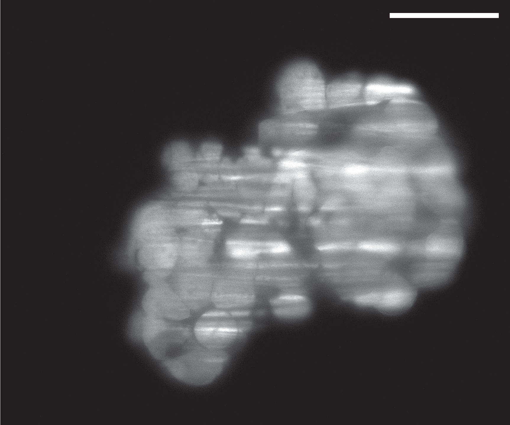
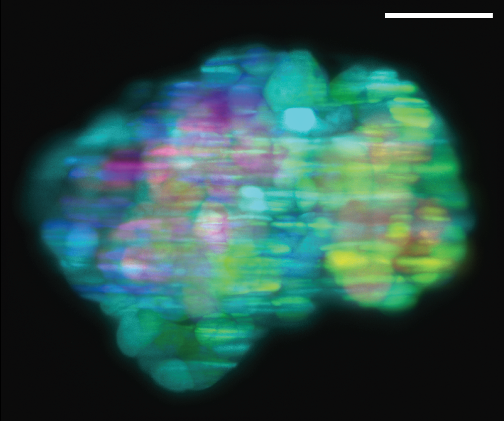
Live spheroid from patient-derived ovarian carcinoma cells transfected with lentivirus containing EGFP construct. Mounted in 1.2% agarose. Z-stacks with 1µm spacing recorded with brightfield and light sheet illumination. Brightfield illumination: white LED, 22ms exposure time. Flat field correction and Gaussian-based stack focuser in Fiji. Light sheet illumination: 491nm laser, band-pass 525/50nm emission filter, 50ms exposure time. Single plane at 40µm into the sample. Depth projection generated using temporal color code with ‘Spectrum’ look-up table in Fiji. Scale bar = 50µm. Sample by Marcin Iwanicki, Stevens Institute of Technology.